폐색전증
정의: 폐 동맥의 혈전색전증
위험인자: 수술 후 움직임 제한, 암, 임신, 약물, 흡연 등
색전 발생 부위: 심부 하지 정맥(m/c), 신장 정맥, 상지 정맥
증상: 갑작스런 호흡곤란, 빈맥, 빈호흡 기침, 흉막성 통증, 객혈
영상소견
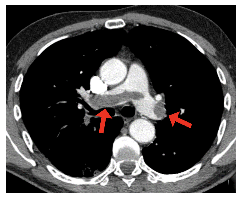
CT : Pulmonary artery 내부의 low density clot 확인
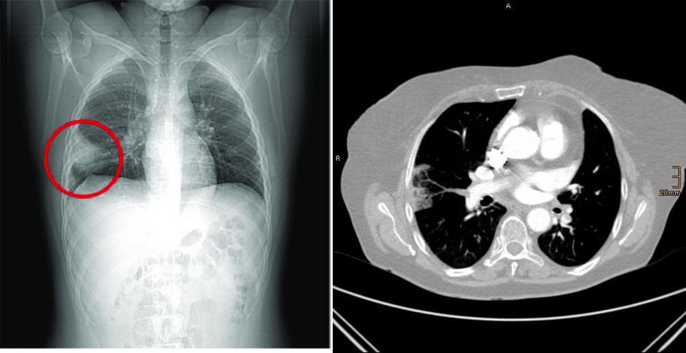
Hampton sign: 혈전색전증으로 인한 폐실질의 wedge-shaped infarction 소견
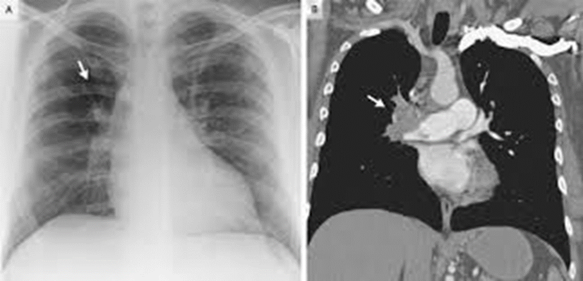
Westermark sign: central pulmonary vessel 확장과 focal peripheral hyperlucency
EKG 소견
sinus tachycardia
new onset atrial arrhthmia
new RBBB
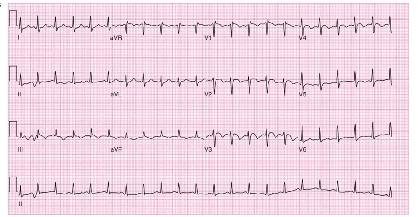
S1Q3T3: Lead I의 prominent S wave, Lead III의 Q wave + T inversion
T wave inversion in V1~V4
예시)
→ S1Q3T3와 함께 V1~V4 T wave inversion이 보이는 소견
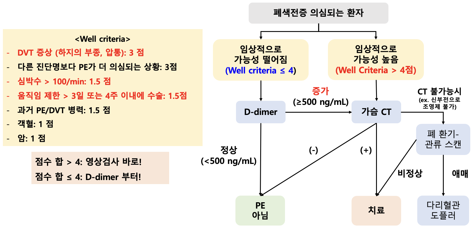
폐색전증 진단
[1] Harrison 21e, pg.2091~2098
[2] UpToDate - Epidemiology and pathogenesis of acute pulmonary embolism in adults, Pathogenesis and Pathophysiology - Risk factors, Source
[3] UpToDate - Clinical presentation, evaluation, and diagnosis of the nonpregnant adult with suspected acute pulmonary embolism, D-dimer, Lower-extremity ultrasound with Doppler
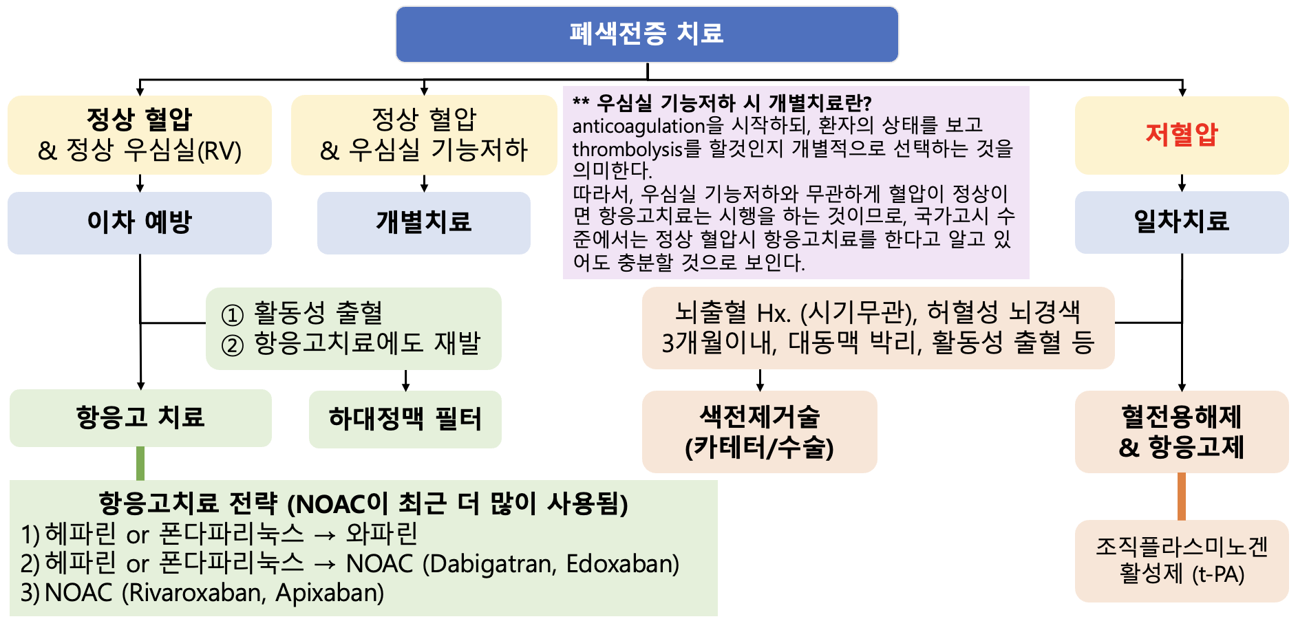
폐색전증 치료
[1] Harrison 21e, pg. 2098~2100
[2] UpToDate - Approach to thrombolytic (fibrinolytic) therapy in acute pulmonary embolism: Patient selection and administration, contraindications to thrombolysis
0개의 글
** 제목만 보더라도 어떤 내용인지 알 수 있도록 완성된 문장으로 작성해주세요.
예시) 초음파 (X) → 초음파 사진에서 PDA 소견을 어떻게 알 수 있나요? (O)