가슴엑스선검사 (CXR)
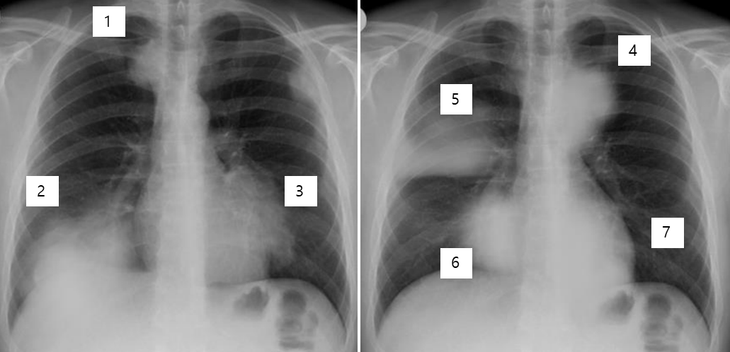
[Normal Anatomy]
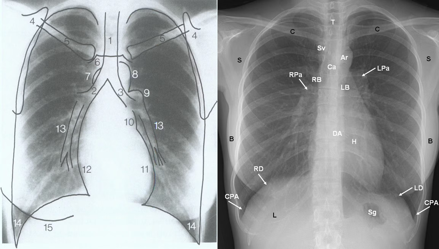
흉벽 (rib, clavicle, scapula, vertebrae), soft tissue
Trachea - carina - Rt/Lt bronchus, Deviation
Hilum (Rt<Lt)
Mediastinum
widening, mass
CT ratio
Heart border (Aortic knob - Lt. pulmonary a., LA appendage, LV apex)
Diaphragm (Rt>Lt, CPA, subdiaphragmatic air/stomach gas, flattening/rising)
Lung field
location, description
Radio-opaque: consolidation, GGO, nodule/mass, reticular opacity, atelectasis
Radiolucent: pneumothorax, emphysema
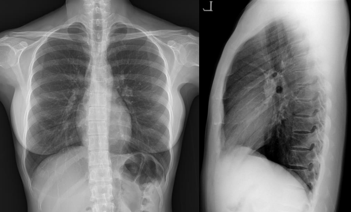
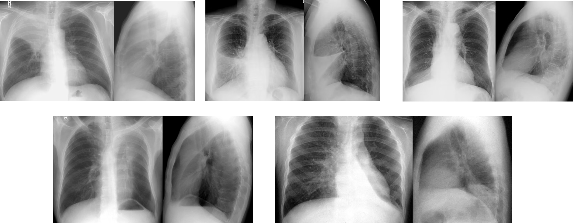
[Normal Chest PA/Lat]
Terminology
Consolidation/Air bronchogram
consolidation: alveolus 내부에 염증, 삼출액, 혈액, 세포 등이 공기를 완전히 대체하면서 폐가 하얗게 보이는 소견
air-bronchogram: consolidation 을 배경으로 endobronchial air가 대비되어 보이는 소견
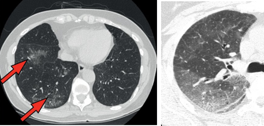
Ground-glass opacity (GGO)
Consolidation보다는 덜 진한 hazy opacity. 폐혈관의 음영을 소실시키지 않는 정도의 음영
염증, 삼출액, 혈액 등으로 폐포강이 부분적으로 채워져 있거나, interstitium의 비후, 폐포의 부분적 허탈 등의 종합적 소견
Ground-glass nodule
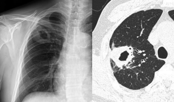
Cavity
consolidation, mass, nodule 내부의 괴사가 기관지를 통해 빠져나가고 그 부위가 공기로 차 있는 형태 (abscess, pneumatocele, TB, cancer)
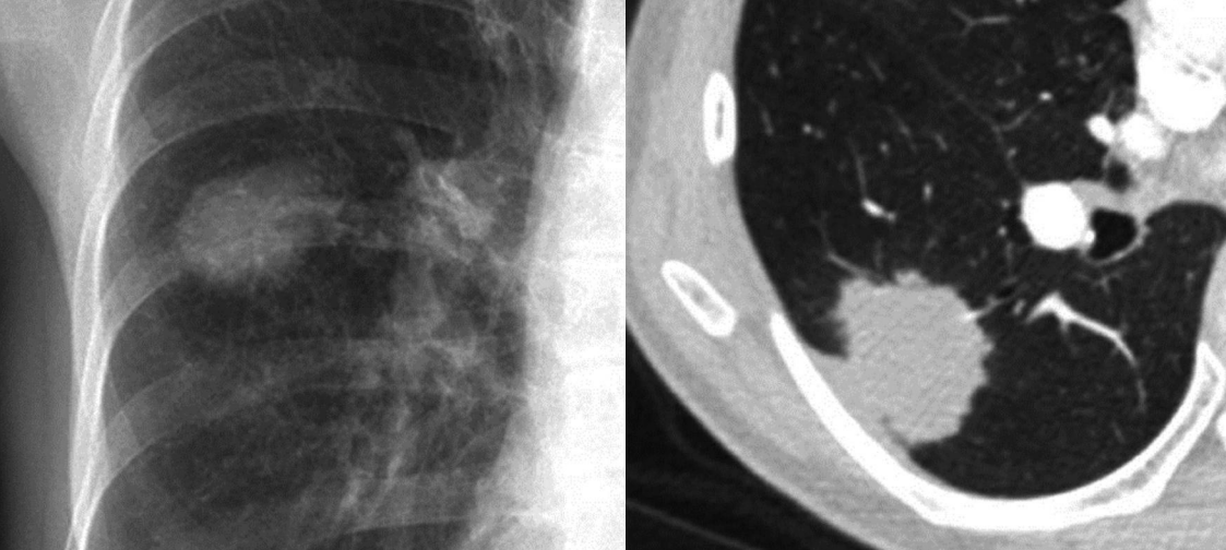
Nodule/Mass
nodule : 3cm 미만의 덩어리 음영
mass: 3cm 이상의 덩어리 음영
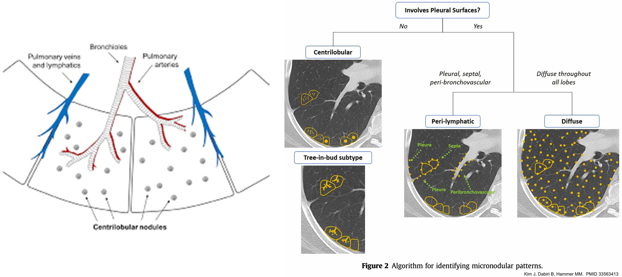
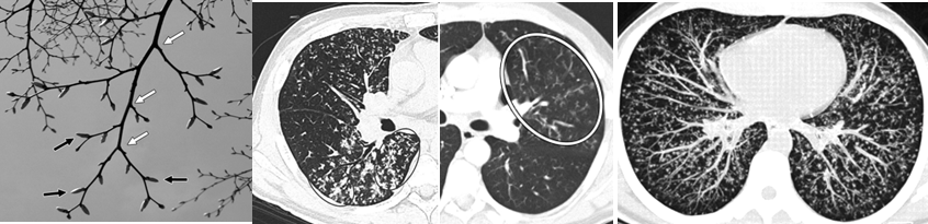
Micronodules
Tree-in-bud: 폐의 중십소엽기관지 내부에 염증, 점액, 액체 등이 차거나 기도벽의 염증성 비후, 확장 등에 의해 나뭇가지가 발아하는 모양처럼 보이는 소견
diffuse panbronchiolitis, tuberculosis 등에서 보이는 소견
Reticular opacity
Interstitial space (interlobular septum)에 fluid가 차거나 fibrosis가 발생하면서 나타나는 음영
ex. Kerley’s B line (pulmonary edema), viral/mycoplasma/PJP pneumonia, sarcoidosis, IPF, lymphatic carcinomatosis 등
Interlobular septal thickening
Silhouette sign
X선 사진에서 음영증가 병변이 심장, 대동맥, 횡격막과 인접해 있을 때 경계면이 소실되어 보이는 소견 (해부학적 연결이 없을 때는 경계가 소실되지 않음)
→ 병변의 위치를 파악하는데 중요한 소견Consolidation, atelectasis, mass 등 공통적으로 해당
Rt paratracheal → RUL
Rt diaphragm → RLL
Lt heart border → LUL, lingula segment
Rt aorta → anterior mediastinum / LUL
Rt chest wall → RUL ant. segment
Rt heart border → RML
Retrocardiac → LLL / posterior mediastinum
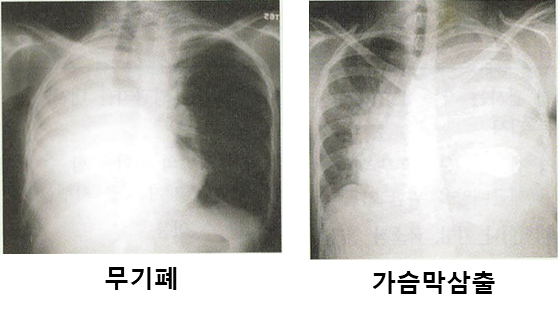
무기폐
경계가 매우 명확한 opacity, air-bronchogram이 없음
Volume loss로 인해 병변쪽으로의 trachea/hilum/diaphragm의 displacement가 발생
Lobar atelectasis
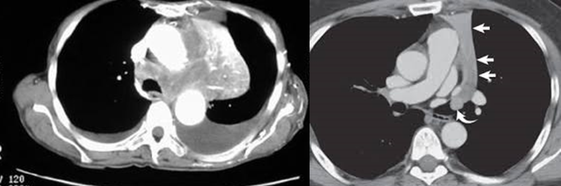
Total atelectasis vs massive pleural effusion
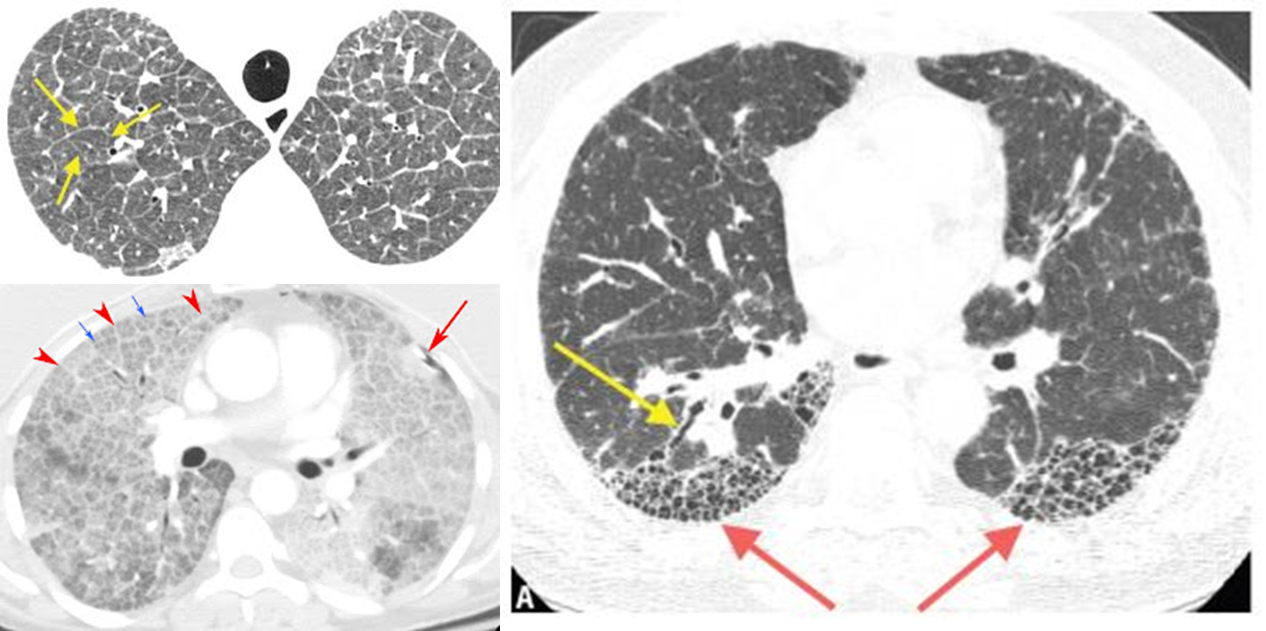
CT 소견
질환 별 요약
각 영상 소견은 각론을 참고
호흡기감염
bronchiolitis
lobar pneumonia
bronchopneumonia
PJP/CMV pneumonia
Lung abscess
septic embolism
결핵
활동성 폐결핵 (Tree-in bud, cavitation, upper lung zone, LN enlargement with central necrosis)
Miliary Tb
결핵 합병증 (aspergilloma, chronic empyema, rasmussen aneurysm)
폐암
Atelectasis, Golden S sign
solitary pulmonary nodule
Hematogenous pulmonary metastasis (“cannonball metastasis”)
Hilar/mediastinal lymph node metastasis
SVC syndrome
pleural metastasis/malignant mesothelioma
Pulmonary edema
interstitial pulmonary edema(kerley’s B line)
hydrostatic pulmonary edema (Bat wing appearance)
permeability pulmonary edema (ARDS)
reexpansion edema
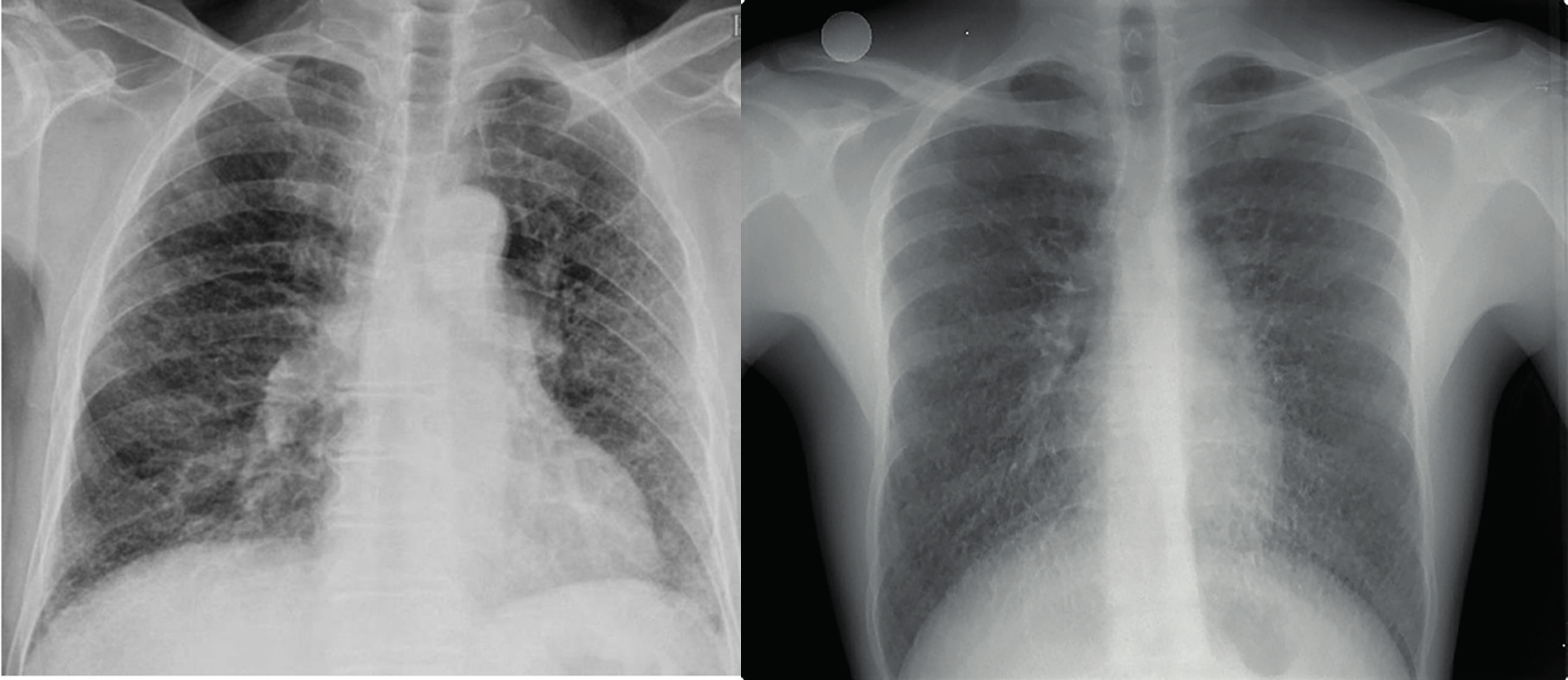
간질성 폐질환
pneumoconiosis (reticulo-nodular, egg-shell calcification, crazy pavings, PMF)
asbestosis (pleural plaque, reticulo-nodular opacity, round atelectasis)
IPF (honeycomb appearance), NSIP, COP
CTD-related ILD, Radiation pneumonitis
sarcoidosis (bilateral hilar lymphadenopathy, reticulonodular opacity)
COPD
폐혈관질환
폐색전증
diffuse alveolar hemorrhage/pulmonary endometriosis
종격동 질환
pneumomediastinum
mediastinal mass
mediastinal widening
흉막, 가로막, 흉벽 질환
흉막삼출
기흉
심혈관계 질환
0개의 글
** 제목만 보더라도 어떤 내용인지 알 수 있도록 완성된 문장으로 작성해주세요.
예시) 초음파 (X) → 초음파 사진에서 PDA 소견을 어떻게 알 수 있나요? (O)